Detection of occult metastases in colon cancer
Metastases pose a great challenge in the treatment of cancer. They are both difficult to detect and to treat. Therefore, the BIOMETSCO team focused on metastases in colon cancer.
"Many colon cancer tumors have already metastasized at the time of diagnosis. And in some cases, the metastases are undetectable by conventional methods. These metastases, known as occult metastases, are our primary focus," says Lasse Sommer Kristensen, who is heading the BIOMETSCO project.
The complex process of metastasis – understanding can lead to better treatments
It’s important to focus on colon cancer. It is the third most common cancer in the world and in Denmark alone, 4000 patients are diagnosed with cancer each year. Although more patients are diagnosed at an earlier stage than before, 20% has already developed metastases at the time of diagnosis. This is bad news. Especially if they are found at distant parts of the body: Only 13% of patients with distant metastases survive 5 years – compared to a 91% 5-year survival rate for colon cancer without metastases.
How the cancer cells spread from the primary tumor to other parts of the body is a very complex process and not well understood. Consequently, the current treatment for metastatic colon cancer is far from optimal. Most colon cancer patients undergo surgery to have the primary tumor removed and many also receive chemotherapy aimed at the metastases. The problem is that the chemotherapy is nonspecific and often causes severe side effects, and it may not necessarily be administered to the right patients. “We must find a better way to identify which patient will benefit from chemotherapy,” says Lasse Sommer Kristensen and adds: “If we can understand more of the complex process of metastasis, we will also learn how the metastases differ from the primary tumor. When we know this, we can identify biomarkers that will allow us to test, whether there are in fact any metastases in a patient.”
Lasse Sommer Kristensen imagines that patient blood samples can be analyzed for the presence of the metastasis-specific biomarkers. If the biomarker is there, so are the metastases. “A better understanding of metastasis can potentially also lead to better metastases-specific treatments with fewer side effects. If specific biological processes are taking place in the metastatic cells, we might be able to prevent the cells from metastasizing,” says Lasse Sommer Kristensen. This pretty much sums up the aim of the BIOMETSCO project: To understand the process of metastasis and thereby identify new biomarkers and potential drug targets.

A two-step method for finding the genes that stand out from the crowd
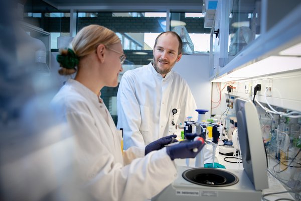
When asked about his approach to identify new biomarkers for the metastatic cells, Lasse Sommer Kristensen modestly replies “We started out as many others have done before. We performed a bulk analysis on two different types of tissues and compared them to see how the expressed genes in the tissues differ.” Basically, the BIOMETSCO team wanted to find out which genes are present in the metastatic tissue but not in healthy or primary tumor tissue. In a bulk analysis, a tissue sample containing different types of cells is analyzed as a whole. A cancer tissue sample therefore contains both the cancer cells themselves but also all sorts of other cell types know as stromal cells. You can think of the bulk tissue samples as a pile of differently colored building blocks with each color representing a different type of cell. The bulk analysis will tell you which genes are expressed by all the different building blocks in the pile. But it’s difficult to tell, which cells express a specific gene. Therefore, the bulk analysis serves only as a first step to identify potentially interesting genes.
The second step in the experimental setup adds more complexity, but Lasse Sommer Kristensen explains that it’s worth the trouble: “Spatial transcriptomics can tell us exactly which cells express a specific gene found in the first step. At the same time, it tells us where the cells are located in an intact tissue sample.” You can imagine the building blocks no longer lying in a messy pile but rather in a very discrete two-dimensional figure with similarly colored block being placed in discrete patterns next to each other. The two-dimensional figure is the intact tissue and the patterns of colored building blocks are specific cell types in the tissue. The spatial transcriptomics method pinpoints exactly which building blocks express a given gene, and where the building block is located in the overall figure. “In our project, this was especially important because we had a ‘prime suspect’ when it comes to metastatic cells in colon cancer,” explains Lasse Sommer Kristensen. The BIOMETSCO team’s ‘prime suspect’ was cancer cells budding off from the main tumor mass at the invasive front of the primary tumor. And the team wanted to know which genes stood out from the crowd of other genes expressed in these budding cells compared to rest of the tumor cell mass.
Biomarker candidates from the invasive tumor front
It’s not only the BIOMETSCO team, who’s interested in the budding cells at the invasive tumor front. This frontmost area of the primary tumor is believed to be the home of aggressive tumor cells that may be responsible for metastasis. “We’ve investigated the invasive front in stage II tumor samples, which by definition, have not yet metastasized to the lymph system or distant sites. However, a significant proportion of these patients have occult liver metastases, and we believe that the invasive front plays a key role in the metastatic process” explains Lasse Sommer Kristensen. The BIOMETSCO team has paid special attention to these invasive fronts by comparing the gene expression at this site with other tissue samples from both tumor mass, liver metastases, and healthy control tissue.
The comparisons were first performed using bulk tissue samples and then followed by spatial transciptomic analysis. This 2-step approach resulted in a list of candidate biomarker genes, which were validated by other methods. The best performing biomarkers were selected for subsequent analysis. “We are looking very much forward to further validate our biomarker candidates. Our biomarker candidates were selected based on a cohort of fresh frozen tumor tissue, but now we have moved to a much larger cohort of well-characterized patients with formalin-fixed paraffin- embedded tissues available,” says Lasse Somme Kristensen. Through the collaboration with Henrik Hager, who works in the clinic, the academic team has access to more than 700 patient samples.
The most important aspect of the larger cohort is not only the number of samples but also the accompanying data. Lasse Sommer Kristensen highlight two very specific advantages of the cohort: “First, we have access to all patient journals. This means that we can make sure that all samples have been categorized correctly as stage 2 tumors. We can also ensure that the patients don’t have hereditary cancer or other types of concurrent cancers. Secondly, the patient samples were collected 5-10 years ago. Thus, patients were treated according to the current guidelines, and we have sufficient clinical follow-up time to categorize patients into those with occult metastasis at the time of diagnosis and those without.
The BIOMETSCO team made a large effort to ensure that the available data was used in the best possible way. Two PhD students manually checked the samples and the accompanying journals. But the project team not only handled large amount of input data. They also generated enormous amounts of data in the project.
The BIOMETSCO project opened new research avenues and collaborations
“Spatial transcriptomics, the second step in our biomarker discovery process, generated a lot of data,” says Lasse Sommer Kristensen. He explains that the BIOMETSCO team zoomed in on the budding cancer cells at the invasive tumor front. “Based on the genes that the different cell types expressed, we divided the cells into different groups. The specific cancer cells within a group were located together, as expected if they evolve and expand based on gaining new advantageous mutations. However, this did not apply to one group of cancer cells, which seemed randomly distributed at a first glance. However, upon a closer look, we realized that these cancer cells included most of the budding cells. And of course we wanted to take a closer look at these cancer cells,” he says. These specific cancer cells also turned out to be located in close proximity to a certain type of fibroblasts, and since it is unlikely that different cells suddenly adapt the same behavior by chance, the BIOMETSCO team reasons that the fibroblasts re-program the cancer cells and make them more prone to metastasize.
Lasse Sommer Kristensen and the rest of the team are keen to learn more about the re-programmed cancer cells and their interactions with the surrounding fibroblasts. “I think we have found something really exciting here. But I must admit that it can be difficult to see the big picture or the relevant patterns, when we are handling these huge amounts of data. Therefore, we’ve teamed up with a team of researchers in Switzerland, who are AI experts,” he explains.
Together with the Swiss researchers, Lasse Sommer Kristensen received a grant from The Danish Cancer Society for the project ‘Investigation of the diagnostic potential of spatial molecular analyses of the tumor microenvironment in colon cancer’ in 2023. This project builds on the findings from the BIOMETSCO project, and in the 3-year project the new research team will use AI approaches to further understand how the local tumor microenvironment influences on the metastatic process. “We are looking forward to taking the work from the BIOMETSCO project a step further. The project has spurred new research avenues, and I hope that the biomarkers, we have identified, will become valuable tools in the clinic in the future,” Lasse Sommer Kristensen concludes.