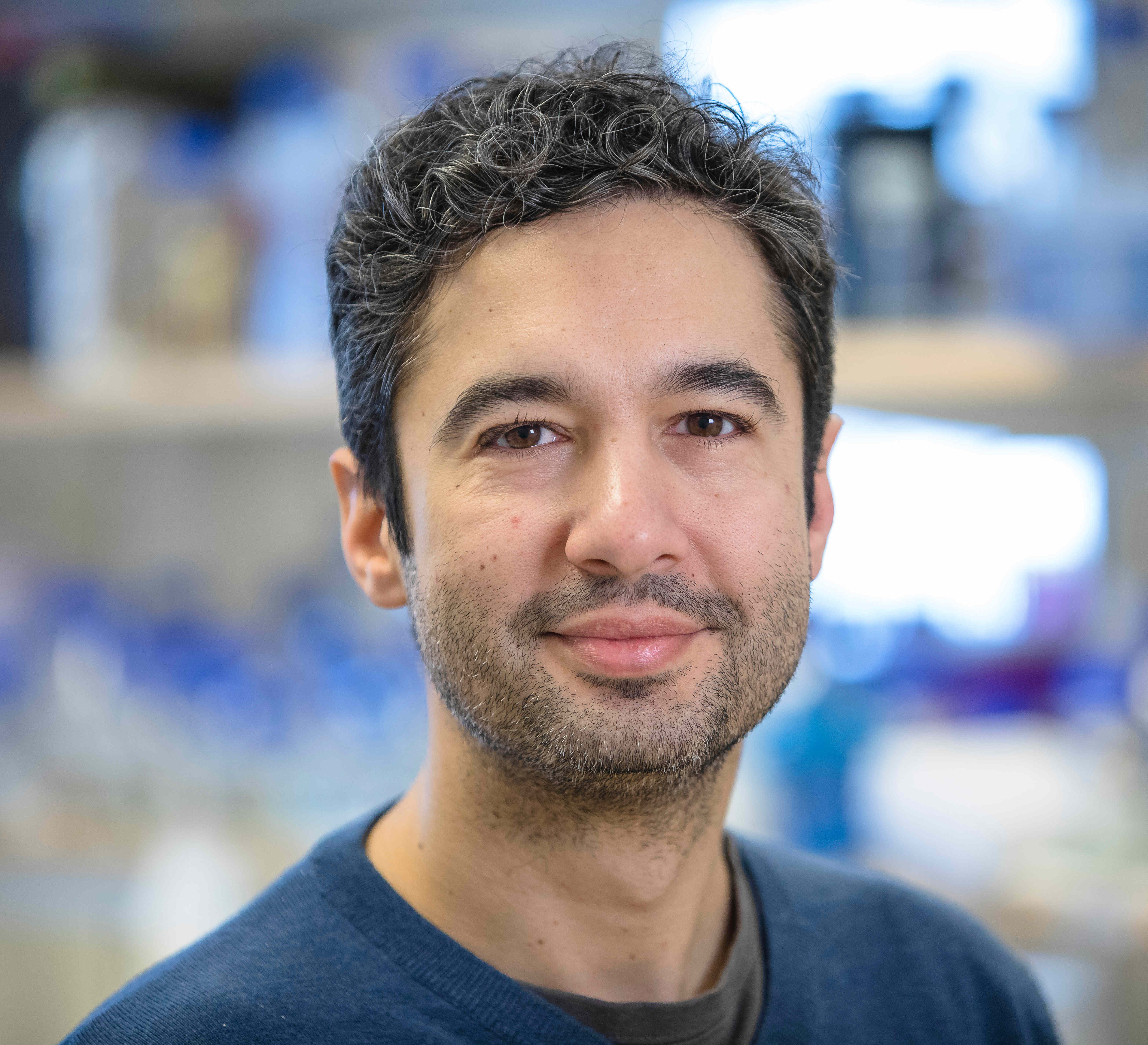
Research Portrait: The Cava lab studies the “Achilles’ heel” of bacteria, the cell wall
Felipe Cava, MIMS Senior Principal Investigator and Associate Professor at the Department of Molecular Biology at Umeå University, wants to understand the diversity of cell wall biology across species and how this knowledge can be used to fight emerging infectious diseases.

Note: This article originally appeared on the MIMS website: https://www.mims.umu.se/news-events/2090-research-portrait-the-cava-lab-studies-the-achilles-heel-of-bacteria-the-cell-wall.html.
Dr. Felipe Cava and his team are using cutting-edge methodology to understand the role of the cell wall in environmental adaptation and for finding potential targets for antimicrobial therapies. The Cava lab and their collaborators recently published two scientific articles in which they revealed groundbreaking discoveries about how the bacterial cell wall adapts to different conditions. The researchers involved in these studies have been able to, for the first time, analyze peptidoglycan, the main cell wall component, in deep details.
New intracellular environment specific mechanism of immune evasion of Salmonella discovered
The environment of the compartment that Salmonella inhabits inside the infected cell triggers changes in its cell wall with consequences on immune signaling. This interference with immune signaling is related to the ability of the pathogen to remain hidden for long periods of time inside the cell.
Bacteria of the Salmonella genus are pathogens responsible for a high percentage of gastrointestinal infections that can sometimes spread to internal organs, posing a risk to human and animal health. Another characteristic of this pathogen is its ability to reduce its growth rate inside the cell it infects. The immune system recognizes structures outside the body itself, one of them being peptidoglycan, the main component of the bacterial cell wall, with recognition receptors both in extracellular fluids and inside cells. However, it is not known how this recognition occurs intracellularly given the scant information on the structure of the peptidoglycan when the bacterium is inside the cell.
Some bacterial pathogens modify their peptidoglycan without the need to perceive certain signals or stimuli, thus reducing their recognition by the immune system, a phenomenon known as immune evasion. A new study co-led by researchers from the lab of Felipe Cava, provides a new insight, identifying modifications in the structure of the Salmonella peptidoglycan that are stimulated once the pathogen has access to the intracellular environment of the cell it infects. This work is the result of collaboration with the groups led by Francisco García del Portillo, CSIC researcher at the National Center of Biotechnology (CNB-CSIC), Ana San Félix at the Institute of Medicinal Chemistry (IQM-CSIC), M. Graciela Pucciarelli and Juan Ayala at the Severo Molecular Biology Center Ochoa (UAM-CSIC).
The results, published in PLoS Pathogens, have been feasible thanks to the application of new ultra-sensitive liquid chromatography and mass spectrometry methodologies that allow obtaining great information from very small amounts of material.
"For the first time, and after the structural analysis of the Salmonellapeptidoglycan when it is located in an intracellular compartment, we have verified that this environment stimulates changes not previously detected in other locations" highlights Francisco García del Portillo, CSIC researcher at the National Center of Biotechnology (CNB-CSIC).
“The analysis of the cell wall has never been done in such a deep manner with such a small amount of sample. This is a fantastic methodological breakthrough with the use of untargeted MS-MS”, says Felipe Cava, researcher at Umeå University and The Laboratory for Molecular Infection Medicine Sweden, MIMS.
Some modifications in the structure of this peptidoglycan, such as a change of the amino acid D-alanine, for alaninol (an amino alcohol) in about 2% of the side chains of the peptidoglycan, reduces the activation of the pro-inflammatory response. The confirmation of anti-inflammatory activity in peptides with this amino alcohol obtained by chemical synthesis highlights the importance of this new finding.
This study opens up new ways of selectively eradicating intracellular infection, proposing the inhibition of alaninol formation as a novel way of acting on infections caused by Salmonella.

Figure: Working model depicting how PG editing in non-proliferating intracellular S. Typhimuriuminterferes with immune signaling and promotes persistence. (A-B) Comparison of structural alterations identified in the PG of intracellular S. Typhimurium versus those reported in other pathogens under laboratory conditions and the structure of a canonical muropeptide of Gram-negative bacteria (extracellular); (C) Scheme that shows how the release from intra-phagosomal S. Typhimurium of outer membrane vesicles having as cargo atypical PG fragments, like those containing D-alaninol, could favor immune evasion and persistence by interfering with NOD1/NOD2 responses. The model compares such event with an infection in which S. Typhimurium releases pro-inflammatory (canonical) PG fragments that stimulate NF-κB. The model also integrates the NOD1 activation by pathogen effectors that target Rho-GTPases independently of PG fragments reported by others [77]. How the PG fragments are transported from the phagosome or vesicles to the cytosol remains poorly understood. NAG: N-acetylglucosamine, NAM: N-acetylmuramic acid, m-DAP: meso-diaminopimelic acid; D-Ser: D-serine, D-Lac: D-lactate, NCDAA: non-canonical D-amino acid; OMV: outer membrane vesicles. Figure is property of the authors of the scientific publication.
Scientific article: SB Hernández, S Castanheira, MG Pucciarelli, JJ Cestero, G Rico-Pérez, A Paradela, JA Ayala, S Velázquez, A San-Felix, F Cava, F García-del Portillo. Peptidoglycan editing in non-proliferating intracellular Salmonella as source of interference with immune signaling. Plos Pathogens 2022; 18(1):e110241; DOI: http://doi.org/10.1371/journal.ppat.1010241
Original press release appeared by CNB-CSIC Communication on 4 February 2022: https://www.cnb.csic.es/index.php/en/science-society/news/item/1937-identificado-un-nuevo-mecanismo-de-evasion-inmune-de-salmonella-especifico-del-ambiente-intracelular
The groundkeepers of the periplasmic space: lytic transglycosylases
Under the lead of Felipe Cava at Umeå University and Tobias Dörr at Cornell University, a big scale study of lytic transglycosylases was carried out to identify the importance of these enzymes in cell wall remodeling. Their findings got published in the scientific journal eLife.
The bacterial cell wall is fundamentally made of peptidoglycan, a mesh-like polymer that surrounds the bacterial cell conferring strength, as well as resistance to internal turgor pressures. Maintaining the integrity of the peptidoglycan sacculus is vital to cell viability however, the peptidoglycan sacculus is not a static structure, rather it is continually expanded and turned over. Lytic transglycosylases are the “space-making” enzymes. They cleave the peptidoglycan to permit its expansion as well as they allow the insertion into the cell wall of protein complexes such as the flagella and secretion systems. If cell wall turn over fails, due to malfunction in the lytic transglycosylase activity, the peptidoglycan biogenesis is interrupted. Big chunks of the peptidoglycan polymer will start to accumulate in the periplasm, resulting in the phenomenon of crowding. Periplasmic crowding is toxic for the cell because it disturbs many intracellular functions. This work sheds light into the unprecedented role of lytic transglycosylases to prevent toxic crowding of the periplasm.
“These enzymes are redundant; they need to make sure that crowding does not happen. We created different types of mutants and multiple knock-outs, to study the function and redundancy of lytic transglycosylases, and we saw that the removal of these enzymes leads to periplasmic crowding. All enzymes we studied are important for building the cell wall, and the cell wall is crucial for the bacteria's’ life. Every fundamental study about understanding how bacteria build the cell wall under different conditions is valuable and could help us to use them as targets”, says Felipe Cava.
Knowledge on these enzymes can open the door to new antimicrobial therapies targeting periplasmic homeostasis, a historically elusive topic.

Figure: Model for lytic transglycosylase (LTG)-mediated removal of toxic peptidoglycan (PG) debris. (A) Endopeptidases (EPs, yellow) excise PG strands (red) from the sacculus (blue), permitting sacculus expansion. (B) In wild-type cells, LTGs (green) digest excised, uncrosslinked PG strands into smaller fragments that can be recycled by AmpG (black) or released through porins (violet). (C) In LTG-deficient cells, excised PG debris crowds the periplasm and becomes toxic. Figure is property of the authors of the scientific publication.
Scientific article: AI Weaver, L Alvarez, KM Rosch, A Ahmed, GS Wang, MS van Nieuwenhze, F Cava, T Dörr. Lytic transglycosylases mitigate periplasmic crowding by degrading soluble cell wall turnover products. eLife 2022;11:e73178 DOI: 10.7554/eLife.73178