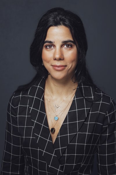
NCMM-EMBL Group Leader Irep Gözen works within chemical life sciences, bionanotechnology and membrane systems
Dr. Irep Gözen has a PhD in chemistry from the Chalmers University of Technology, Sweden, and was previously a postdoctoral researcher in Professor Vinothan Manoharan’s group at Harvard University. She joined NCMM as head of the Bionanotechnology and Membrane Systems Group in 2016. She also holds an adjunct associate professor position at the Department of Chemistry, University of Oslo.

Can you tell me about your research in a nutshell?
We specialize in the materials aspects of complex biological problems; focusing on the substance excluding the genetic polymers, proteins and chemical energy compounds of the cell. These primitive structures consisting mainly of biological membranes, can still perform many of the cellular functions, even migration, and can teach us about the potential role of membranes in complex biological behavior. By bringing membranes in contact with surfaces, we aim to develop an artificial cell model programmable by environmental cues such as temperature, as well as to form and functionalize the intracellular units of cells and most recently to discover new features of protocells, the primitive containers which are thought to have given rise to life.
Can you elaborate on your recently published article, “A Nanotube-Mediated Path to Protocell Formation,” and describe how your group worked together on the project?
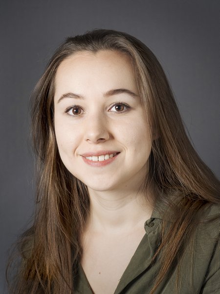
Elif Köksal, PhD fellow from Gözen’s group comments on the article, published recently in ACS Nano: “Our project is about the emergence of life on earth. In this article, we report on a new way for the formation of first primitive cells, so called ‘the protocells’, and point to the role of surfaces in protocell formation, growth and reproduction.”
In a majority of studies investigating protocells, the researchers have focused on compartments formed in solution. By adding mineral surfaces to the bulk solution, we see an entirely new way of vesicle formation. We found that when we add phospholipids to a solution on top of a mineral-like surface, protocells connected via a network of nanotubes allowing their communication, spontaneously form. In our paper, we describe how exactly the mineral-assisted protocells are able form and grow and also separate from the population when a gentle flow is applied. We also talk about the potential implications of this new formation pathway for the currently open, highly debated questions such as protocell division.
How is your latest research approached by other scientific experts?
We have so far received a quite positive response. We presented our work at the annual meeting of the American Biophysical Society early this year. The Society highlighted our findings with an interview included in their press package which was later picked up by other news sites from all over the world. On a separate occasion, we had the privilege to show our results to Professor Jack W. Szostak from Harvard Medical School, who is one of the pioneering researchers when it comes to the origin of life. It was great to get his feedback and encouragement. I look forward to seeing what response we will get from a wider audience now that our article is online.
What do your findings show and what do you think this research will lead to?
We are currently working on entirely mimicking the early Earth environment. We have so far used phospholipids in our studies. Many believe that fatty acids came first because they are simpler molecules. We are trying to simplify the molecular structure of the ingredients and want to show that fatty acids can form similar compartments on mineral surfaces. At the moment our protocells do not contain genetic material. Elif is currently working on incorporating RNA fragments into our protocells. We want to investigate if these compartments can share the encapsulated material within the network and see if/how the genetic material can be encapsulated and redistributed. These findings can change the way protocell formation, growth and reproduction have been assumed to occur.
Do you have any thoughts on the next steps for using this model? What applications (translational or otherwise) do you think this could be used for?
Our research is currently focusing on answering a basic scientific question in the context of the origin of life. From a technical perspective, our method is the only one other than microfluidics-based methods, which can consistently produce precisely unilamellar vesicles (vesicles with a single lamella) in numbers as high as 20 million per a standard microscope cover slip. While microfluidic-based techniques require special equipment and expertise, the experiment we show can be conveniently reproduced without the need of special infrastructure. Unilemallar vesicles are used for investigation in many areas of research, for example to study the behavior of membrane proteins.
Can you tell me about your research group?
Elif Köksal is currently working on the project described above. Another PhD fellow Karolina Spustova is investigating compartmentalization and how different reactors form within the cell. Our research assistant Gizem Karabiyik is working on developing a protocol for harvesting nanoparticle assemblies which spontaneously form by sensing curvatures on soft materials. One other PhD fellow Aysu Kubowicz works on membrane damage and investigates how biological membranes fracture as observed in earthquakes. We also have two postdocs: Inga Põldsalu is an expert in ink-jet printing technology and can make polymer-based actuators. Lauri Viitala who knows a great deal about photo-thermally programmable phospholipids, is also a part of my group, but he is located at Chalmers University of Technology in Sweden where I have a visiting position since 2016. We are in the process of recruiting one more PhD student. My group is growing and it is great to have this opportunity to interact with another university outside of Oslo.
Who do you currently collaborate with?
We have many national and international collaborators: We work with Prof. Michael Taylor from Santa Clara University, USA, for the development of computational models which can explain the mechanical aspects of membrane behavior in detail. Prof. Aldo Jesorka from Chalmers University is another international collaborator working on microfluidics integrated surface acoustic wave sensors, highly useful tools for sensitive detection of lipid membrane-surface interactions. Prof. Paul Dommersnes from NTNU, a theoretical physicist highly knowledgeable in membrane biophysics, is a close national collaborator. Most recently, we started collaboration with Prof. Henrik Friis from the Museum of Natural History to be able to get more insight on natural mineral surfaces.
Are you still involved in the UiO: Life Science convergence environment initiative? How is that progressing?
In our convergence environments project, supported by the UiO: Life Sciences, we have a consortium consisting of four principal investigators. This is an ongoing, highly interdisciplinary project and is a way to combine different expertise to tackle major challenges within health and environment. The first part of the project was to make the cell-like compartments, which we have now achieved. Associate Professor Andreas Carlson’s group from the UiO Department of Mathematics developed the mathematical model for the above-mentioned protocell studies, to explain why the compartment formation is favored. Next is the functionalization of the compartments together with Professor Harald Stenmark’s group at the Institute of Clinical Medicine, University of Oslo.
In parallel we work with Prof. Gry Oftedal from the Faculty of Humanities, UiO, a philosopher of science working on the concept of scientific metaphors and investigating whether they facilitate the public’s understanding of scientific concepts or if it builds barriers. We use terminologies in our research, such as ‘artificial cells’ or ‘cell-like constructs’ which potentially could confuse a non-scientific audience. Currently, together with Gry and Ingrid Lossius Falkum from the Department of Philosophy, UiO, we are preparing a pilot study to determine the intuitive understanding of scientific metaphors. It is important for us to combine various skills to characterize the phenomena we observe in the laboratory as well as to communicate it to the public and study on metaphors is really an enrichment to the project.
- Visit https://www.softlabnorway.com/ for more information about the Gözen Group
- Visit https://pubs.acs.org/doi/10.1021/acsnano.9b01646 to read the article, “A Nanotube-Mediated Path to Protocell Formation”.
- Visit https://www.uio.no/english/research/strategic-research-areas/life-science/research/convergence-environments/ for more information on the UiO convergence environments.