Meet BICU: a research infrastructure and environment for bioimaging in Sweden
Meet BICU Director and MIMS Team Leader Richard Lundmark and BICU Facility Manager Irene Martinez Carrasco, and learn about BICU’s expertise and approaches in light and correlative microscopy and in the study of biomolecule affinities.


The Biochemical Imaging Centre Umeå (BICU) is a research infrastructure and community that offers a variety of state-of-the-art imaging and biomolecule affinity technologies for the local, national and international research communities.
Supported by The Laboratory for Molecular Infection Medicine Sweden (MIMS), the Swedish node of the Nordic EMBL Partnership, the interdisciplinary facility is hosted by the Department of Medical Biochemistry and Biophysics in the Chemical Biology Centre (KBC) at Umeå University in Sweden.
Heading BICU is Richard Lundmark, a MIMS Team Leader and Professor at the Department of Integrative Medical Biology at Umeå University. Prof. Lundmark directs and coordinates BICU’s development along with Facility Manager Dr. Irene Martinez Carrasco. Prof. Lundmark explains BICU in a nutshell: “We provide a smörgåsbord of light microscopy instruments together with instruments that help people to measure interactions between biomolecules together with very competent personnel that help the research get done.”
The seeding of BICU began a little over 10 years ago when Prof. Lundmark and some colleagues opened up access to a few of their microscopes. He explains that “at that time, BICU wasn't funded in any way; it was simply us providing microscopes to people who wanted to use them.”
In 2014, BICU recruited Dr. Martinez Carrasco, a research scientist from Spain with experience working in a microscopy core facility. Prof. Lundmark describes Dr. Martinez Carrasco’s impact and how the center has become what it is today:
We were very happy to recruit Irene! She came to us with knowledge about how to run the facility, and she is brilliant at it. Then we got new microscopes, and it took off from there. We continue to evolve with the researchers using the facility. We have PhD students, postdocs, and other researchers using all of the instruments, and they tell us what they need, how they need it to be adapted, what could be improved, and so on. BICU very much depends on the scientific environment. Thanks to Irene, we have an environment that is about the users: what can be done and how can we improve together?
According to Dr. Martinez Carrasco, it’s the bottom-up approach that allows BICU to meet the scientific needs: “Richard has a lot of contact with PI's, and I have regular contact with users who tell us what they need. That is our main goal - to facilitate the use of the technology.”
Scientific research at Umeå University is steeped in infection biology expertise with strengths in biochemistry, as well. According to Prof. Lundmark, it’s primarily these areas that drive the imaging technology development at BICU. Dr. Martinez Carrasco gives examples of projects:
We have about 50 research groups, mostly from Umeå, using the instruments. Our international users are typically connected to a local group. We get a lot of infection projects. For example, users may want to determine localization of their proteins or visualize and study interactions. They look at effector molecules and how bacteria and viruses invade cells.
New developments in advanced imaging technologies are underway at BICU. For example, working together with the Umeå Center for Electron Microscopy (UCEM), BICU is driving the development and application of correlative light-electron microscopy (CLEM) in Sweden. CLEM enables high-resolution imaging by electron microscopy of the same samples viewed by live cell fluorescence imaging. This approach couples the power of the two techniques to provide integrated images of high resolution areas of interest in a cell or tissue sample.
Prof. Lundmark explains additional developments at BICU:
We're also focusing a lot on being able to measure interactions in living cells using light microscopy. It complements our capabilities in affinity measurements nicely. In addition, we are working on the fast imaging of processes with SIM (structured illumination microscopy). SIM is a super-resolution imaging technique that also allows for high temporal resolution of biological processes in living samples.
Capabilities and expertise at BICU
BICU offers instrumentation, project consultation, training and technical support for image and data acquisition and analysis in a wide array of light microscopy and affinity measurement techniques.
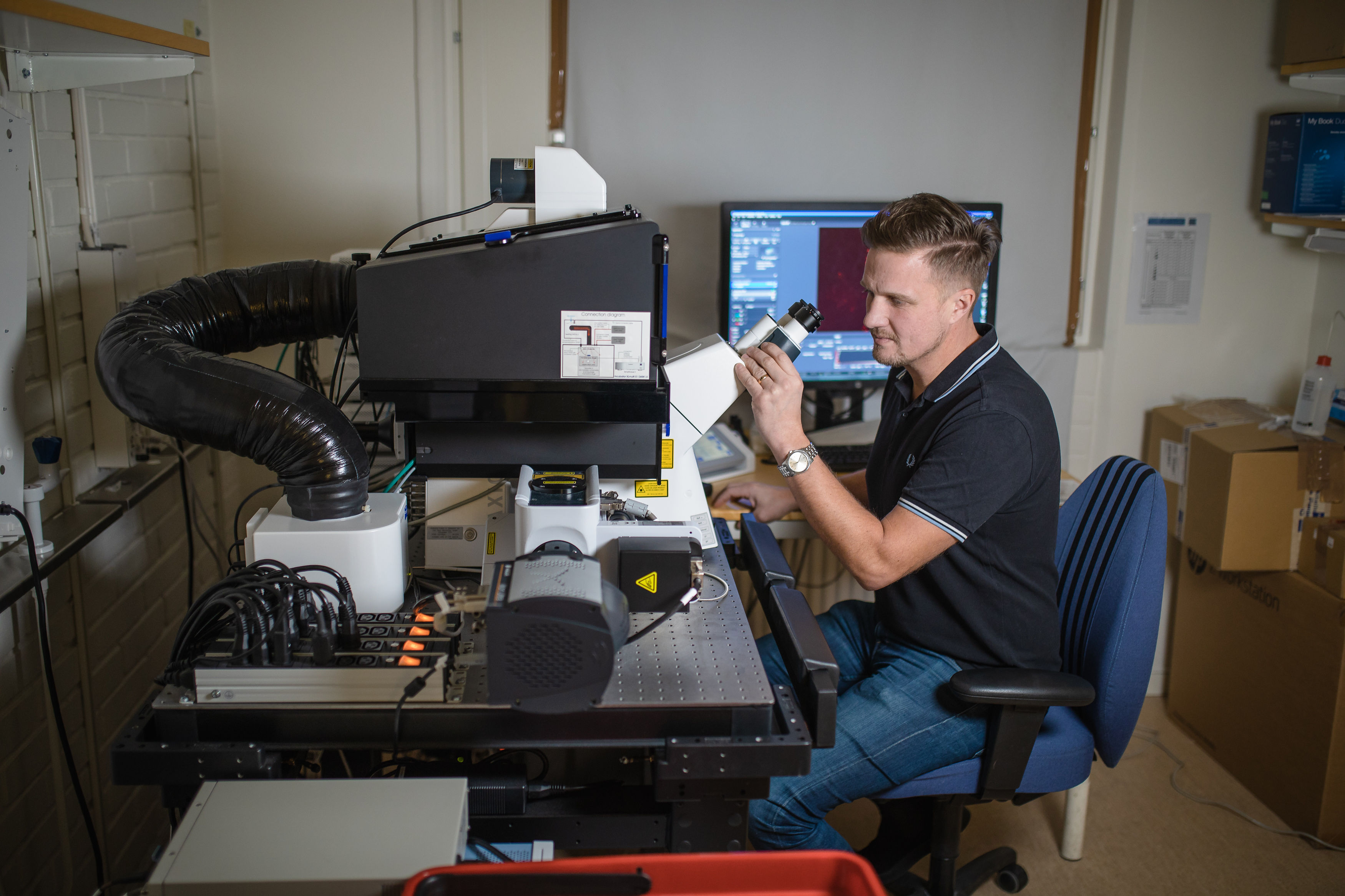
There are eight light microscopes for live cell imaging, including some super-resolution approaches: FLIM (fluorescence-lifetime imaging), FRAP (fluorescence recovery after photobleaching), FLIM-FRET (fluorescence resonance energy transfer), TIRF (Total internal reflection fluorescence), and spinning disk and 4D confocal. The up-to-date inventory of instruments can be found at the BICU website. The facility also offers correlative imaging with two different microscopy approaches, e.g., CLEM in collaboration with UCEM.
For biomolecule (protein, RNA, lipid, carbohydrate) affinity studies, BICU houses seven different instruments for measurements in solution and on a polar or nonpolar surface, including Biacore 3000 for biomolecule interactions, Proteon XPR36 for bacterial binding studies, Auto-Isothermal Calorimetry for biomolecule interactions in solution, LigandTracer Green for study of interactions in real-time, CD spectrometer for protein confirmation studies, and Quartz Crystal Microbalance with Dissipation monitoring (QCM-D) for analysis of lipid membrane binding.
Atomic force microscopy is also available for examining protein-protein interactions and their geometry and strength at the nano and pico-newton scales.
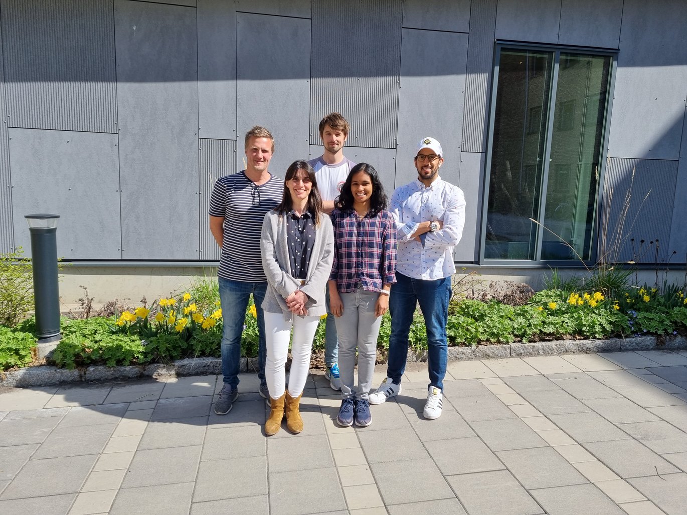
The power of a microscopy facility, as opposed to simply a collection of instruments, is that technical expertise is available. Two experts, Gayathri Vegesna and Johan Olofsson Edlund, round out the facility team along with Prof. Lundmark as director and Dr. Martinez Carrasco as manager and expert in light microscopy. Prof. Lundmark explains the complementary competences of Drs. Vegesna and Olofsson Edlund:
Gayathri came to us from Rainer Pepperkok’s group at EMBL-Heidelberg. She works with both electron and light microscopy and is the correlative microscopy expert, shared between BICU and UCEM. She trains and supports users on both light microscopes and electron microscopes.
And:
Johan is our affinity instrument expert. He's only been with us for less than two years, but we can already see the impact - a significant growth in the usage of these instruments because of the support he can give. It just took the right person to come in. They're blooming now.
BICU is set up as a decentralized facility with super-users in addition to the facility staff. This set-up allows for a dynamic and flexible environment that can respond quickly, with the appropriate expertise, to needs such as BSL2 work and adoption of new technology.
Dr. Martinez Carrasco and Prof. Lundmark both acknowledge the importance of users and super-users in the wide array of competences in the BICU environment. Dr. Martinez Carrasco explains: “With this flexible organization we rely a lot on the users; they take very good care of the instruments.”
She continues from the practical point of view of user training and instrument management: “With the pandemic we have implemented online training, and we can also remotely log-in to instrument computers, making the management of the satellites easier.”
National and international networks in bioimaging
To bring CLEM to users, particularly in Sweden, BICU works very closely with UCEM, the Umeå Center for Electron Microscopy, also affiliated with MIMS and at Umeå University.
BICU and UCEM together form one of the five nodes of the Swedish National Microscopy Infrastructure (NMI), providing national expertise in correlative light-electron microscopy.
Funded by the Swedish Research Council, the NMI was founded with nodes of expertise in 3D correlative multimodal imaging (University of Gothenburg), super-resolution fluorescence microscopy for nanoscale biological visualization and high-throughput multiplexed profiling (KTH Royal Institute of Technology), intravital animal imaging with multi- and single-photon microscopy (Stockholm University), and correlative light-electron microscopy (Umeå University). A fifth node was recently added to provide expertise in bioimage informatics (Uppsala University). Dr. Martinez Carrasco explains:
The network covers the most challenging and latest imaging technologies. We cannot know or do everything in one single facility, but together, across the country, we have the expertise. We collaborate and offer courses together. And, our users can initiate projects at the different nodes.
Prof. Lundmark adds:
The staff at each node come together in a super structure for light microscopy in Sweden, keeping the country at the forefront. And we use the other nodes. For example, we don't have a STED microscope (Stimulated Emission Depletion) here in Umeå, but in Stockholm they are focused on this technique and have great competence to which we can direct users.
In the Nordic region, BICU has contacts with imaging specialists in Finland and Denmark, and participates in the new Bridging Nordic Microscopy Infrastructure network. On the European scale, BICU interacts with the imaging experts at EMBL and within the Euro-Bioimaging European-wide research infrastructure, primarily via the Swedish NMI.
People and community are at the core
Dr. Martinez Carrasco explains what brought her to BICU and what motivates her toward service in science:
I’m here because I like people. I like to interact with the users and help them. I started in a facility as a user and really liked what they were doing. Then I ended up working there. I have liked science and biology since I was small. I had my first microscope when I was 16, a real one!
She continues:
This is a job where you are constantly with people. You need to like being around them. We may teach users how to use an instrument, but importantly we answer their questions and support their research. They need to feel like they can just come to us for help. We can try different technologies, techniques, or instruments together. A lot of people may give up without the support we can offer. And, we learn from all of the users. That’s so important also.
Prof. Lundmark remarks on his path to imaging:
I’ve always liked visual concepts. I think in pictures. During my PhD, I had to borrow a microscope. It was a fluorescence microscope, but it was in the middle of a bright lab. So I had to use a big rain coat to cover it, and myself, while I worked! I liked the microscopy approach and I’ve also always liked science. I'm inspired by the fact that you can have a hypothesis, and if you design the experiment right, you can answer yes or no. I also had a microscope as a kid!
Building a research infrastructure that scientists want to use and contribute actively to, in just a few years, is no small feat. Dr. Martinez Carrasco reflects on this challenge and accomplishment: “I didn't expect the facility to grow as much as it has by this point. The biggest challenge with a facility is building trust with the users and PIs. Over time, day-by-day, user-by-user it has come to being the community it is.”
Prof. Lundmark adds:
I'm proud that we’ve been able to construct a research environment, not just a facility. Many research groups, together with the facility staff, solve scientific problems. I want to be able to address more scientific questions, and in order to do that, I need help from other researchers both to think about how, but also to actually get the instruments we need.
A research core facility can be simply a service-station that provides infrastructure for science or it can also become a community, as Prof. Lundmark sums it up: “the instruments are essential, but it’s the people that drive and use them that really facilitate the science. It’s the people that make the community.”
Bioimaging and affinity measurement for your research project
The Biochemical Imaging Centre Umeå (BICU) at the Department of Medical Biochemistry and Biophysics in the Chemical Biology Centre (KBC) at Umeå University offers expertise, consultation, instrumentation, training and technical support for image and data acquisition and analysis as well as laboratory space and cell culture facilities.
This national center for advanced fluorescence super-resolution and live cell imaging is funded by Umeå University and the Swedish Research Council, as a part of the National Microscopy Infrastructure (NMI) of Sweden, providing support for personnel and laboratories. Researchers apply for university and/or external funding for new instruments. User fees support the running costs of the instruments. BICU and the Umeå Centre for Electron Microscopy are a node in Sweden’s National Microscopy Infrastructure (NMI) for correlative microscopy.
Researchers interested in using the BICU facility or consulting with the staff about projects are welcome to contact the BICU staff.
Mobility funding from a NordForsk Infrastructure Hub grant may help cover costs for visits to the facility for researchers working at Nordic EMBL Partnership institutes. Please contact your local administration for more information.
With support from NordForsk, MIMS is organizing a course in correlative microscopy for Nordic researchers 5-9 December 2022. Follow updates on the program and registration.
The “Technologies Advancing Molecular Medicine” series highlights the people and activities in research technologies and core facilities in the Nordic EMBL Partnership for Molecular Medicine.
CONTACT
Richard Lundmark
Director, Head and Coordinator
richard.lundmark@umu.se
Irene Martinez Carrasco
Facility Manager
irene.martinez@umu.se