November 2020

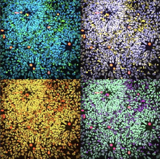
A Compilation of the Rosette-forming Neural Progenitor, by Safak Coglayan (overall winner), NCMM
"In our group we aim to understand how our brain is developed using a neuronal differentiation protocol that broadly models the fundamentals of this process. The picture is a pop-art visualization of the rosette-forming neural progenitor cells that are derived from human embryonic stem cells. Cells are labeled with antibodies to stain paired box protein PAX6 and phosphorylated form of histone H3 protein. Nuclear labeling is achieved with a DNA binding fluorescent stain, DAPI. I take the pictures using Zeiss LSM 800 confocal laser scanning microscope and process them using Zeiss ZEN Blue software."