June 2020
Nano Violet, by Aysu Kucukturhan, NCMM
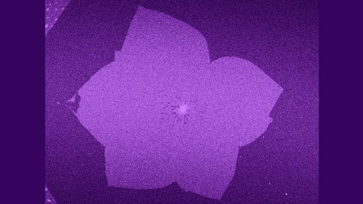
"The image shows the confocal microscopy scan of two stacked lipid bilayers from top view, the upper one of which has spontaneously ruptured and evolved to a shape reminiscent of a flower. The thickness of each of the lipid layers is approximately 5 nm."
"For my PhD project, I am investigating the ruptures occurring in biomembranes under mechanical tension. To form the membranes, I deposit micron-size spherical lipid reservoirs onto planar solid surfaces, the bright spot at the center that looks like the pistil of the ‘flower’. The reservoirs spontaneously spread on the surface in 2D and transform into double bilayers. While the lower layer continuously adheres onto the surface, the expansion of the upper bilayer causes a buildup in membrane tension and eventually, the spontaneous rupturing."
"The ruptures occur in all kinds of interesting morphologies, among them: a violet."