February 2020
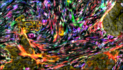
Colourful Microenvironment, by Teijo S Pellinen, FIMM
"Clear cell ovarian carcinoma stained with multiplexed immunohistochemistry (in-house technology at FIMM; Blom et al 2017 Scientific Reports). The colourful cells are fibroblasts (four different fibroblast markers, PDGFRA, PDGFRB, FAP, aSMA) and the yellow colour shows the tumour cells. White/gray are nuclei. The name of the image could be: The colourful tumour-microenvironment of clear cell ovarian carcinoma."