February 2019
Cancer Fish, by Susanne Hultsch, FIMM
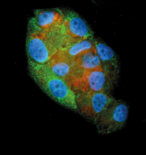
"The image was taken with the Nikon 90i microscope fluorescent microscope. It is a 40x image of a tamoxifen-resistant breast cancer cell line (T-47D Tam1). In blue is the nucleus, and in green is Ceruloplasmin (CP), a ferroxidase enzyme. This was highly up-regulated in the tamoxifen-resistant cells and is very useful as a cytoplasmic marker. In red we have Lysosomal-associated membrane protein 1 (Lamp1) and as the name indicates it is a marker for lysosomes. The lysosomes in those cells have an abnormal phenotype and contribute to the tamoxifen resistance.
"The image came from testing some different staining parameters. In this case, I tested the permeabilization of the cells with Triton-X, which on one hand makes epitopes for the antibodies (LAMP1 and CP) better accessible, but it destroys the lipids in the cell. It was too aggressive to depict the accumulation of lipids in the lysosmes, so that those images with this setting never made it to the publication (https://bmccancer.biomedcentral.com/track/pdf/10.1186/s12885-018-4757-z). The ones with Filipin, an alternative to Triton-X that also stains cholesterol, were able to make it, because this kept the lysosomes intact."