Advances in stem cell engineering prove that changes in mechanics contribute to cell physiology. However, it remains a challenge to understand the mechanisms by which cells sense and respond to mechanical signals from the extracellular environment. JVN has previously invented a nanoporous biomaterial that can be controlled in its microstructural layout. Thereby it became possible to modulate the mechanical environment around single cells growing on or within the material. Pilot experiments showed that material stiffness influences gene expression; a result that confirms existing mechanobiological theory.
Recent research suggests that the geometry of extracellular environment also plays an important role for cell regulation. We will use JVN's biomaterials to verify this hypothesis, and make it more explicit. We aim at developing a mathematical model that allow us to identify important morphological traits of the material microstructure.
To this end, we will develop and apply methods from stereology, stochastic geometry and biomechanics to data. We collect 3D images of cells cultured in geometrically different nanostructures, captured by synchrotron generated X-Ray tomography, as well as high resolution 2D electron microscope images. This 3D and 2D information together will be used to fit a stochastic model for the geometry of the biomaterial, a "digital twin". The digital twin will allow us to simulate the environment of cells that live in the pores of the material. We will use mechanobiological models to calculate the forces that cells experience, and predict their reaction.
Our project combines methods from various fields of science (click on + to read more):
Mechanobiology studies how living cells are influenced by mechanical forces and seek to link force fields to changes in expressions of biomolecules. This relatively new field of science got a lot of attention when a seminal experiment showed that the development of stem cells into specialized cells such as neurons or osteoblasts depends on the stiffness of the material they were growing on. Previously, cell differentiation was thought to be exclusively controlled by chemical cues.
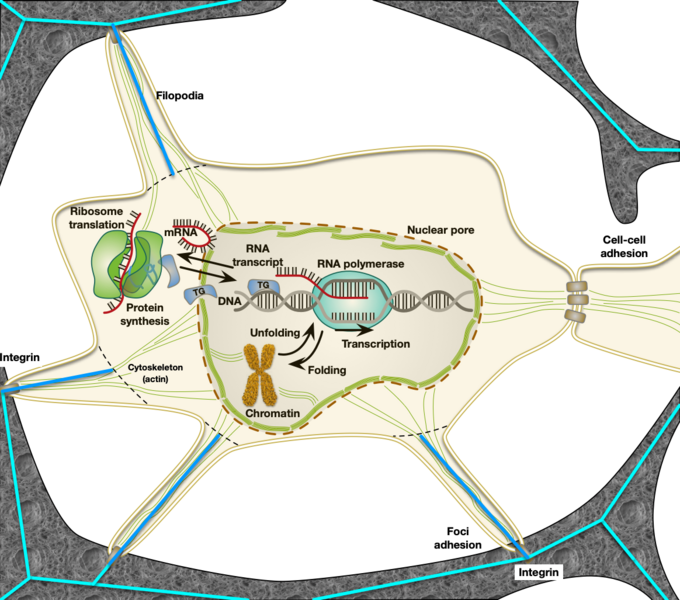
The picture symbolizes a cell located in the cavity of a biomaterial, how force propagate between a cell and its environment through structural elements, and how intracellular processes that extract genome information are affected.
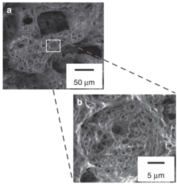
In this project, we will have access to high resolution 3D X-ray tomography images from the material. Still, these images do not represent the small pores adequately. We will therefore combine 3D information with easier accessible 2D transmission electron images. To this end, we will need to stereology: a collection of methods to quantify 3D geometric properties, e.g. of materials or biological tissue, when only images of sections are available. Stereological methods comprise special sampling techniques and statistical estimators for 3D quantities such as volume fraction, surface area, or pore size diameters.
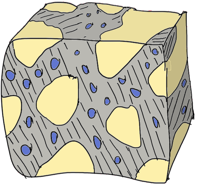
The sketch illustrates material that has pores of vastly different size. Small pores are underrepresented on 2D sections, since large pores are more likely to be hist by a random plane section through the material. Finding the correct size distribution is known as "Wicksell's problem" in the stereological literature, and it has fuelled mathematical ideas for about a century. Our project gives ample opportunity to formulate and - hopefully - solve similar challenging problems.
Developments in materials science allow us to prepare biomaterials with very specific properties. We work with microstructural designs that can be described as open foams. The material contains pores of various sizes that can be varied. Large pores allow cells to enter, smaller pores can house nanoparticles for drug delivery, [Andersen, Nygaard, Burns, Raarup, Howard, Nyengaard, Besenbacher, Kassem, Kjems, siRNA nanoparticle functionalized scaffolds enables controlled multi-lineage differentiation of human mesenchymal stem cells, Molecular Therapy, 2010:18 (11) 2018-2027.]
With advances in microscopy it has become possible to extract 3D information of the smallest (< 20 nm) structural elements in these materials. We have used synchrotron generated X-Ray tomography, as well as high resolution electron microscope images to reveal the layout of these materials, both with and without the presence of cells. The data from these investigations are used in further mathematical analysis. We focus on descriptions of the force fields present and the statistical variations seen in the structures.
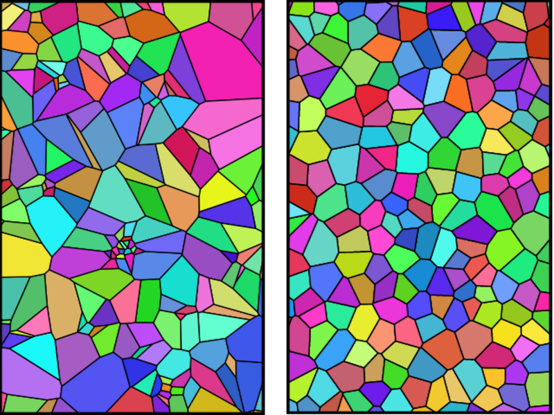
Stochastic Geometry studies random spatial patterns and quantifies their properties. The most important class of models are random point patterns; they can serve as the basis for more complex models such as random tessellations, depicted above. Tessellations can be used to model foam materials.
The figure shows realizations from a so called Voronoi tessellation, a relatively simple model that results when "germ points" grow simultaneously in all directions. The cells stop growing where they touch each other. In the left picture, the germs clustered, while in the right picture, they were quite regularly spaced. Colors are random. In our project, we will use more elaborated models for random tessellations to describe the material's microstructure.
