Unraveling the centromere
Centromeres are essential for healthy cell division. Researchers now reveal the mechanism that allow them to form.

Article originally published at NCMM website: https://www.med.uio.no/ncmm/english/news-and-events/news/2024/unraveling-the-centromere.html
At the Centre for Molecular Medicine Norway (NCMM), researchers are working on defining the molecular components of the centromere, a specialized region within chromosomes. Centromeres are critical for making sure that when a cell divides, the new daughter cells receive one copy of each chromosome.
Without a functional centromere, duplicated chromosomes would be randomly distributed to the daughter cells. Cells with abnormal numbers of chromosomes are usually destroyed. In some cases, however, they may survive, and the presence of these cells is usually associated with diseases such as cancer.
But how does the centromere differ from the rest of the chromosome? And what factors ensure that only one centromere is present and functional on each chromosome?
In a recent study published in Nature Communications, researchers from the Sekulic group at NCMM and collaborators unravel the function of key players that build the centromere.
CENP-A and CENP-B cooperate to form the centromere
The centromere is made up of DNA wrapped around specific proteins, which include CENP-A and CENP-B. While CENP-A is known to be an essential part of the centromere, the role of CENP-B has so far been an enigma.
In this study, the Sekulic group and collaborators at the Swiss Federal Institute of Technology in Lausanne and Osaka University, shed light on the role of CENP-B.
In addition, they uncover in detail how CENP-A and CENP-B cooperate to define and form the centromere.
– In chromosomes, DNA is normally tightly packed in protein-DNA complexes known as chromatin. This makes the DNA relatively inaccessible, explains Nikolina Sekulic, group leader at NCMM.
– We found that presence of CENP-A loosens up the DNA packaging, allowing CENP-B to access and bind to the DNA. Once CENP-B binds, it loosens up the DNA packaging even more. We believe that this “loose chromatin” is very favorable and necessary for the centromere to form, says Sekulic.
– Our results suggest that the cooperative “unpacking” of DNA by CENP-A and CENP-B is required to establish a robust and functional centromere,adds Ahmad Ali-Ahmad, researcher in the Sekulic group.
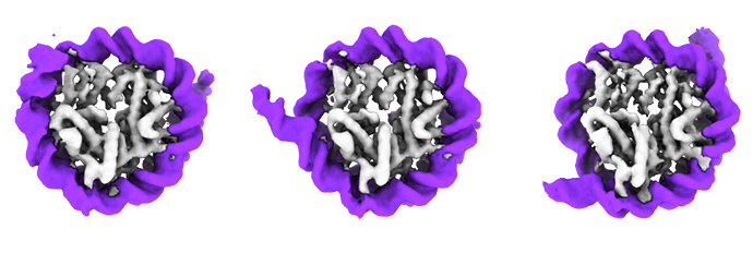
In chromatin, DNA (shown in purple) is wrapped around a protein core (grey). CENP-B (not shown) binds the DNA and induces unwrapping of DNA from the protein core. Credit: Ahmad Ali-Ahmad (Sekulic group).
Findings may explain loss of Y chromosome
Mistakes in chromosome distribution during cell division can happen from time to time. The older we get, the more cell divisions our cells go through, making it more likely for mistakes to occur.
Curiously, the male Y chromosome is particularly prone to becoming lost with age. Recent studies have revealed that such loss of the Y chromosome can be linked to disease development in men, including more aggressive forms of cancers.
The male Y chromosome is the only human chromosome that does not contain CENP-B in its centromere.
Based on their findings of CENP-B, Sekulic speculates that this could cause the Y chromosome to have a less robust centromere. In turn, this could lead to more frequent mistakes in its distribution and subsequent loss over time.
Collaborative effort revealed the role of CENP-B
Demonstating how CENP-A and CENP-B collaborate to form a centromere, was itself a result of a collaborative effort.
Ali-Ahmad from the Sekulic group had observed the effect of CENP-B on DNA using cryo-electron microscopy (CryoEM). CryoEM is a powerful method that allows researchers to visualize protein structures down to the atomic scale.
At the same time, Harsh Nagpal from Fierz' lab in Switzerland had observed a similar effect using a different method, known as single-molecule fluorescence (smFRET) looking at the chromatin fibers.
– It was reassuring for both groups that we were observing the same phenomenon using complementary methods, says Sekulic.
– This collaborative study clearly demonstrates the power of evolving technologies in solving longstanding and important questions. The tools to conduct this study only became available a few years ago with the development of CryoEM and sophisticated chromatin assembling techniques in combination with smFRET, says Sekulic.
Publication
Nagpal, H., Ali-Ahmad, A., Hirano, Y., Cai, W., Halic, M., Fukagawa, T., Sekulić, N., & Fierz, B. (2023). CENP-A and CENP-B collaborate to create an open centromeric chromatin state. Nature communications, 14(1), 8227.