Cryo-EM in Aarhus: from the resolution revolution to in situ structural biology
Meet DANDRITE Affiliated Researcher and structural biologist Thomas Boesen to learn about the power of cryo-EM and the national EMBION Cryo-EM Facility he manages at Aarhus University.

Seeing is believing! Science becomes more accessible when visualized. Demonstrated more clearly than ever during the COVID-19 pandemic, the application of structural biology enables understanding of biological processes in cells and viruses.
Cryogenic electron microscopy (cryo-EM), a technology that uses biological materials trapped in their native state in amorphous ice, has created a revolution in 3D protein structure determination. An atomic resolution view into the structure and dynamics of macromolecules such as viruses and protein complexes, is possible. And, as access to the equipment and computational methods has grown, it is more feasible than ever for researchers to apply the technology to their scientific questions.
At Aarhus University in Denmark, the EMBION Cryo-EM Facility offers state-of-the-art equipment, workflows and support to researchers in academia and industry for studies of biological samples.
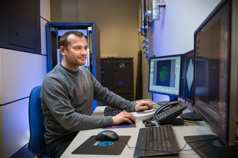
Dr. Thomas Boesen is the Manager of the facility, and Affiliated Researcher at the Danish Institute for Translational Neuroscience (DANDRITE).
A national network in the heart of nanoscience
Aarhus University is one of the founding members and the current host of EMBION, the national cryo-EM facility in Denmark established in 2019. EMBION is a consortium of university partners supported by the Danish Ministry of Higher Education and Science to establish and operate a national cryo-EM facility. The University of Copenhagen serves as the current co-host.
EMBION is open to academic and industry scientists in Denmark. International researchers can also gain access to the platform through specific programs, such as the iNEXT Discovery Program or international partnerships, such as the Nordic EMBL Partnership for Molecular Medicine.
The Interdisciplinary Nanoscience Center iNANO at Aarhus University hosts the EMBION facility. Dr. Boesen explains:
iNANO is a multidisciplinary research and education unit, set up in 2002. It includes groups in chemistry, mathematics, biology, medicine, and physics with a strong commitment to collaborating across fields. The interdisciplinarity in iNANO requires an outstanding research equipment platform for any application from microscopy to diffraction to spectroscopy, easing access to important research infrastructure.
The EMBION Cryo-EM Facility is part of this iNANO research equipment platform, along with a material science EM facility with both scanning and transmission electron microscopes.
A game changer in the resolution revolution
Even before cryo-EM was able to provide high resolution structures, researchers could produce low resolution results of large multiprotein complexes, typically difficult to crystallize. Using this low resolution information, for example by docking crystal structures of subunits, cryo-EM was still a very useful tool for understanding large molecular machines. Originally trained as an X-ray crystallographer, Dr. Boesen contrasts low resolution cryo-EM with crystallography, “with crystallography, if you don't get to around 3Å resolution you cannot really use it for anything, making a strong point for cryo-EM.”
However:
If I consider single particle analysis, I think we've seen the power of cryo-EM ever since the resolution revolution 10 years ago, where developments in the hardware simply made high resolution studies of individual molecules possible. Cryo-EM may soon take over the numbers when it comes to structures deposited in the Protein Data Bank.
Membrane proteins, which make up the majority of drug targets, provide a clear example of this power in cryo-EM technology. First, they can be studied in an environment similar to their native cellular membranes. Second, high resolution information about complexes and protein-protein interactions can be gleaned, as Dr. Boesen explains:
G protein-coupled receptors have been an important structural biology and drug target for many years. Around 15 years ago, the first crystal structures came out, but people had cut pieces off the proteins and added fusions, changing the native state to be able to crystallize them. Although that gave the first structures, progress was slow. But nowadays, high resolution structures of entire receptors, bound to G-proteins, one after another, are being solved. The bottleneck of crystallization is gone. Cryo-EM has been a game changer!
In recent years, Dr. Boesen has been collaborating closely with Prof. Poul Nissen and his research group at DANDRITE determining structures of lipid flippases and the insulin receptor, all transmembrane proteins. In fact, these were some of the first results with cryo-EM to come out of Aarhus.
Cryo-EM as a springboard to in situ structural biology and beyond
Cryo-EM is relatively new in Denmark, compared to neighboring Sweden and Germany. For this reason:
A lot of what we do is to help people get started, as we don't yet have many well-established research groups that have been doing cryo-EM for many years. We focused initially on setting up single particle analysis, and the facility offers training in all aspects of the experimental workflow,
explains Dr. Boesen.
With the single particle workflow well established, Dr. Boesen and his team are setting their sights on new pipelines, e.g., cryo-electron tomography (cryo-ET), a technique of sequential imaging of a target specimen rotated on an axis. Dr. Boesen explains:
High resolution, even a resolution allowing atomic modeling, of macromolecules in their native environment, in situ structural biology, is where we want to go. We have established all the equipment, and are validating workflows so that users can soon get started.
The EMBION team in Aarhus is also setting up microcrystal electron diffraction (microED), a technique that uses nano-sized crystals of small molecule drugs or proteins, often not suitable for X-ray diffraction. Dr. Boesen explains the impact, “the NIH declared microED the Breakthrough of the Year in 2018. We have a large crystallography community here in Aarhus, and microED will be immensely useful here.”
Cryo-EM is available in Aarhus, not just for life science, but also for materials science applications. Dr. Boesen explains the collaboration between the two, “there's really good local synergy between life and material science electron microscopy, for example with very sensitive biological or radiation-sensitive materials. With EM, we can solve structures, but also do elemental analysis.”
There are also applications with light microscopy. Dr. Boesen expounds on such path forward:
We have started collaborating with the bioimaging facility in Aarhus, and we are looking into combining super-resolution microscopy with EM. For example, with correlative light-electron microscopy, you get images from fluorescence light microscopy and from high resolution EM at the same time, and with the right setup combing live imaging and instant high pressure freezing you can get information from a specific time point of a biological process across scales.
Dr. Boesen continues:
I think this is where the future lies. We discuss frequently and visit with colleagues at the Umeå Center for Electron Microscopy to exchange knowledge and be at the forefront of developments. These visits are thanks to support from CryoNET, offering us many interesting interaction points.
Single-cell technologies, e.g., proteomics and genomics, are also advantageous for combining with high resolution imaging, “Normally, when you're looking at cells, you're looking at the average of a population of cells, but what is the role of the individual cells or cell types when you have, e.g., a mix of differentiated cells in tissues and organs”, asks Dr. Boesen. Thanks to support from the Novo Nordisk Foundation, CryoNet will offer a course in the combination of single-cell methods and cryo-EM in Aarhus in spring 2023.
From crystallography to living wires
Dr. Boesen splits his time between running the EMBION Cryo-EM Facility, along with three application specialists and a technician, and leading a mid-sized research group of postdocs and PhD and undergraduate students. Affiliated with the facility team is also an electron microscopy engineer, and working in close collaboration is a scientific computing expert, who is setting up processing pipelines, handling the local computing cluster and managing the data storage clusters. While these teams are indeed separate, Dr. Boesen emphasizes the importance of a tight relationship between research and research infrastructure.
Research interests took Dr. Boesen from crystallography into cryo-EM about 15 years ago. “Combining high resolution crystallography and low resolution cryo-EM was already possible at that point. I learned cryo-EM to use it in this bigger context,” says Dr. Boesen.
These days, Dr. Boesen has let his research interests take him to applying structural biology to cable bacteria in the seafloor in collaboration with the Center for Electromicrobiology:
There are filamentous bacteria that are found in the top few centimeters of water sediments. Imagine that you have 10s of 1000s of cells that are connected end-to-end, forming a filament. It turns out that these filaments make two redox half reactions at opposite ends. Electrons that come from oxidation of hydrogen sulfide deep in the sediment, are transported through this bacterial filament and delivered to reduce oxygen at the top of the sediment, making a living electrical wire.
He continues:
The more we learn about it, the more interesting it gets. It turns out that these bacteria have an internal wire network that is located in the periplasm. And this periplasm is shared through all the cells in a filament.
Purifying the fiber network and studying its conductivity are challenging, but can be done and demonstrate that the source is very conductive. When asked what’s next, Dr. Boesen amazes at the high conductivity and draws on the power of what he knows well:
We want to solve the molecular structure to understand the mechanism of electron transport and how it is coupled to metabolism. How does this wire couple up to enzymes that can up-and download electrons for metabolic redox processes? We'll be combining crystallography and microED with single particle cryo-EM, cryo-ET and in situ structure analysis.
Having leveraged the resolution revolution from a decade ago Dr. Boesen is now looking at the next potential revolution, one he sees could be a combination of single-cell ‘omics and high resolution microscopy, including cryo-EM.
Cryo-EM for your research project
The EMBION Cryo-EM Facility at Aarhus University is a national cryo-EM core facility for researchers within and outside of Denmark. Thanks to continued investments from the Danish Ministry of Higher Education and Science and private foundations such as Novo Nordisk Fonden and Lundbeckfonden, the network provides access to state-of-the-art instrumentation, thorough training, and project support through user fees or collaboration. In addition, facility time can be allocated for users from industry.
Dr. Boesen and his facility team are dedicated to getting users started and exploring if cryo-EM is feasible and beneficial for their project.
Researchers interested in applying cryo-EM technology to their studies can view the instrumentation and workflows available at the EMBION Cryo-EM website. Potential users should contact Cryo-EM Facility Manager Thomas Boesen (thb@mbg.au.dk) or complete the online contact form on the website.
Mobility funding from the Nordic EMBL Partnership Hub NordForsk grant may help cover costs for visits to the facility for researchers affiliated with Nordic EMBL Partnership institutes. Please contact your local administration for more information.
CONTACT
Thomas Boesen
Cryo-EM Facility Manager
thb@mbg.au.dk
The “Technologies Advancing Molecular Medicine” series highlights the people and activities in research technologies and core facilities in the Nordic EMBL Partnership for Molecular Medicine.